Trigger Points and the Sacroiliac Joint

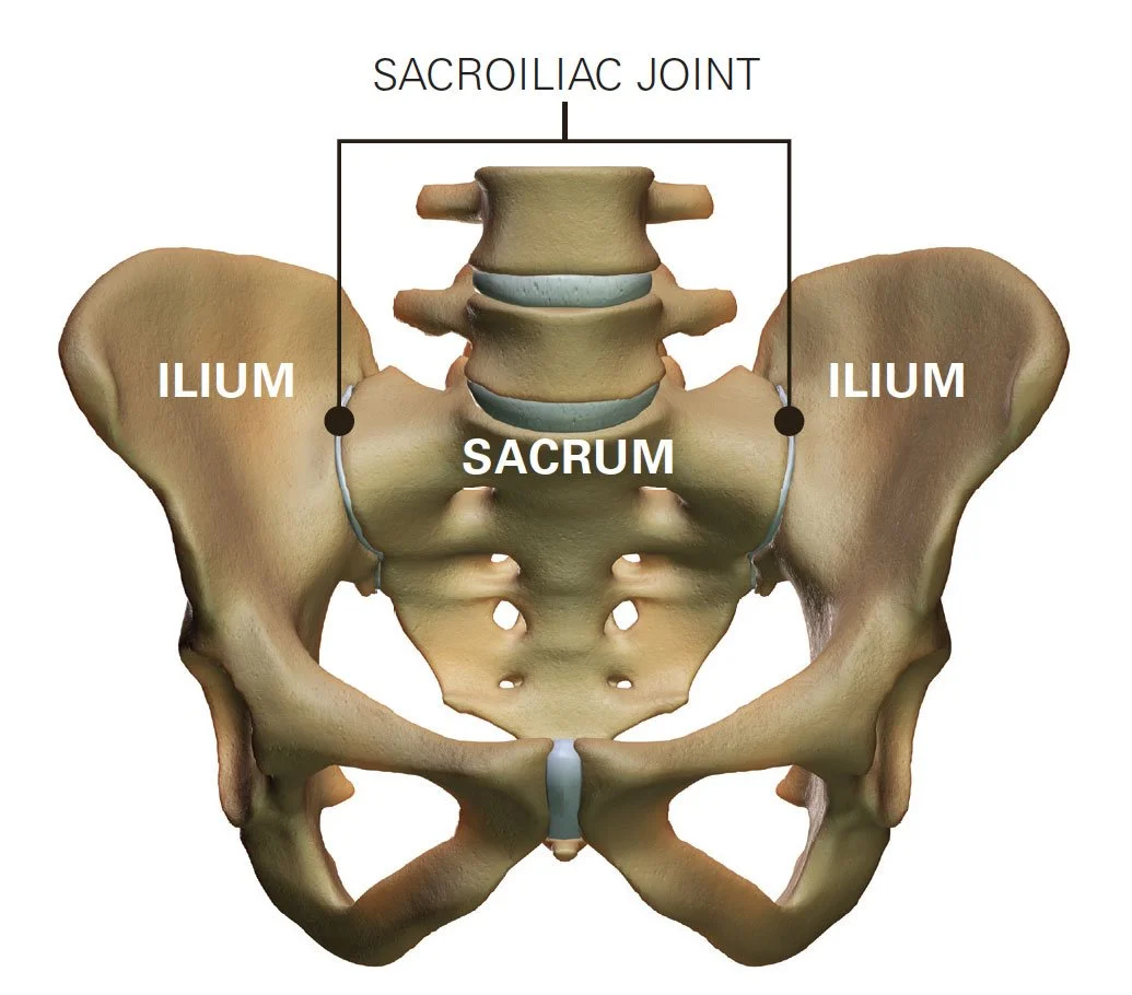
The sacroiliac joint, often abbreviated as SI joint, is a joint in the pelvis that connects the sacrum (the triangular bone at the base of the spine) to the ilium (one of the bones of the pelvis). It plays a crucial role in transferring the weight of the upper body to the pelvis and legs.

The SI joint is unique because it has limited mobility compared to other joints in the body. Its primary function is to provide stability to the spine and pelvis, rather than allowing for a wide range of motion. However, even though it has limited mobility, the SI joint can still be a source of pain and discomfort for some individuals, often due to conditions like inflammation, injury, or arthritis.

Issues with the sacroiliac joint can cause lower back pain, hip pain, and even radiating pain down the legs. Diagnosis and treatment of SI joint problems may involve physical therapy, pain medication, injections, or in some cases, surgical intervention. If you're experiencing pain or discomfort in this area, it's advisable to consult a healthcare professional for an accurate diagnosis and appropriate treatment.

While it is not as extensively studied as more common musculoskeletal conditions, here are some general points about the epidemiology of SI joint dysfunction:

1.     Prevalence: SI joint dysfunction is relatively common but can be challenging to diagnose accurately. The prevalence of SI joint dysfunction varies in different populations, but it is estimated to affect approximately 15-30% of patients with chronic lower back pain.

2.     Age and Gender: SI joint dysfunction can affect individuals of all ages, but it is more commonly seen in adults. It does not appear to have a strong gender predilection, meaning it can affect both men and women.

3.     Underdiagnosis: SI joint dysfunction is often underdiagnosed because its symptoms can be similar to other musculoskeletal or spinal conditions. This can make it challenging to determine the exact prevalence in the general population.

4.     Causes: The causes of SI joint dysfunction can vary and may include trauma, pregnancy-related changes, inflammatory conditions, and degenerative processes. In some cases, the exact cause may not be easily identified.

5.     Comorbidities: SI joint dysfunction may be associated with other musculoskeletal conditions, such as lumbar spine issues or hip problems, which can further complicate diagnosis and treatment.

6.     Diagnostic Challenges: Accurate diagnosis of SI joint dysfunction can be challenging, and it often involves a combination of clinical evaluation, medical history, imaging studies (e.g., X-rays, MRI, or CT scans), and diagnostic injections. Some patients may undergo multiple tests and evaluations before a definitive diagnosis is made.

7.     Management: The management of SI joint dysfunction can include conservative treatments such as physical therapy, pain medications, and injections of corticosteroids or local anesthetics. In some cases, surgical procedures, such as SI joint fusion, may be considered for patients who do not respond to conservative treatments.

It's important to note that the epidemiological data related to SI joint dysfunction is continually evolving as medical understanding and diagnostic techniques improve.


When a physical therapist examines the sacroiliac joint (SI joint) for dysfunction or pain, they typically follow a systematic approach to evaluate the patient's condition. The examination aims to assess the joint's mobility, stability, and the presence of any related musculoskeletal issues. Here are some of the common steps involved in a physical therapist's examination of the SI joint:

Medical History: The physical therapist will start by taking a detailed medical history. They will ask questions about your symptoms, any previous injuries, surgeries, or medical conditions that might be relevant. This information helps them better understand your specific situation.

Subjective Assessment: You will be asked to describe your symptoms, including the location, intensity, duration, and any activities or positions that exacerbate or alleviate your pain. This information helps the physical therapist in forming an initial hypothesis.

Physical Examination:

    • Observation: The therapist may observe your posture, gait (walking pattern), and any visible signs of muscle imbalances or joint asymmetry.

    • Palpation: The physical therapist may use their hands to palpate (touch and feel) the area around the SI joint, looking for tenderness, swelling, or muscle tightness.

    • Range of Motion: They will assess the range of motion of the lumbar spine and hips, as restrictions or limitations can contribute to SI joint issues.

Special Tests: Physical therapists may perform specific orthopedic tests to help diagnose SI joint dysfunction. These tests may include:

    • Gaenslen's Test

    • Thigh Thrust Test

    • Compression Test

    • Distraction Test

    • Sacral Thrust Test

    • Fortin Finger Test

    • SI Joint Compression Test

Functional Assessment: The therapist may assess your functional abilities, including activities such as walking, standing, sitting, and bending. They will look for movements or positions that reproduce your pain.

Muscle Testing: Evaluation of the strength and flexibility of the muscles surrounding the SI joint is important. Weak or imbalanced muscles can contribute to SI joint dysfunction.

Neurological Assessment: The therapist may check for any neurological symptoms or signs related to the SI joint, such as sensory changes or reflex abnormalities.

Radiological and Imaging Studies: In most cases, the physical therapist review results of X-rays, MRI, or other imaging studies to correlate with their clinical examination.

Surface palpation of the sacroiliac joint (SI joint) can be a useful component of the diagnostic process, but its reliability in isolation is limited. Palpation involves manually examining the area around the SI joint for tenderness, swelling, or other signs of discomfort. While it can provide valuable information, several factors can affect its reliability:

--Variability among Individuals: SI joint anatomy and the location of pain or    discomfort can vary from person to person. Some individuals may have naturally tender or sensitive areas around the SI joint, making palpation more subjective.

--Patient Tolerance: The reliability of surface palpation can also depend on the patient's pain tolerance. Some individuals may be more sensitive to pressure, while others may not feel tenderness even when there is an issue with the SI joint.

--Surrounding Musculature: Muscles and soft tissues surrounding the SI joint can also affect the reliability of palpation. Muscle tension or trigger points in the area may cause discomfort that is not directly related to the SI joint itself.

--Experience of the Examiner: The reliability of palpation can be influenced by the skill and experience of the healthcare provider conducting the examination. A trained and experienced clinician may be more adept at identifying subtle signs of SI joint dysfunction.

--Diagnostic Support: Palpation is often used in conjunction with other diagnostic  methods, such as orthopedic tests, imaging studies, and medical history. These additional tools can help confirm the findings from surface palpation.

--Differential Diagnosis: Lower back and hip pain can have various causes, including issues with the lumbar spine, hip joint, and muscles. It can be challenging to attribute pain solely to the SI joint based on palpation alone.

Given these factors, surface palpation of the SI joint is typically considered a part of the overall diagnostic process, but not the sole determinant of SI joint dysfunction. In summary, while surface palpation of bony landmarks is a part of the diagnostic process for SI joint dysfunction, it is typically not considered a standalone or highly reliable method for diagnosis.

The clinicians at Lanier Therapy in Motion use a combination of clinical assessment, orthopedic tests, imaging studies (such as X-rays or MRI), and the patient's history to arrive at a more accurate diagnosis.

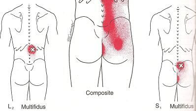
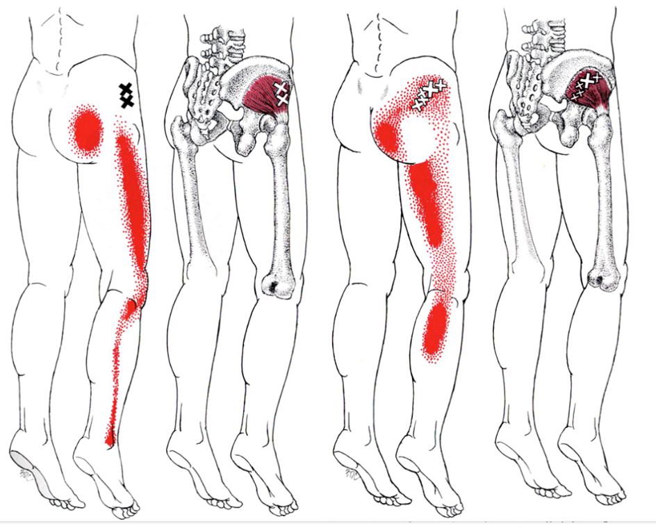
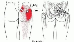
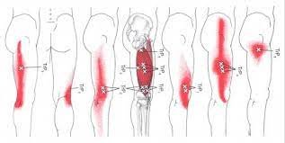
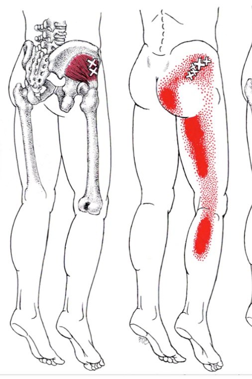
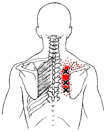
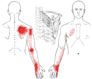
In addition, our clinicians will check for the presence of trigger points in the lower back and sacroiliac joint musculature.  While trigger points themselves do not directly cause sacroiliac joint dysfunction, they can contribute to or exacerbate pain in the lower back, including the region around the sacroiliac joint (SI joint). Here's how trigger points can be related to SI joint dysfunction:

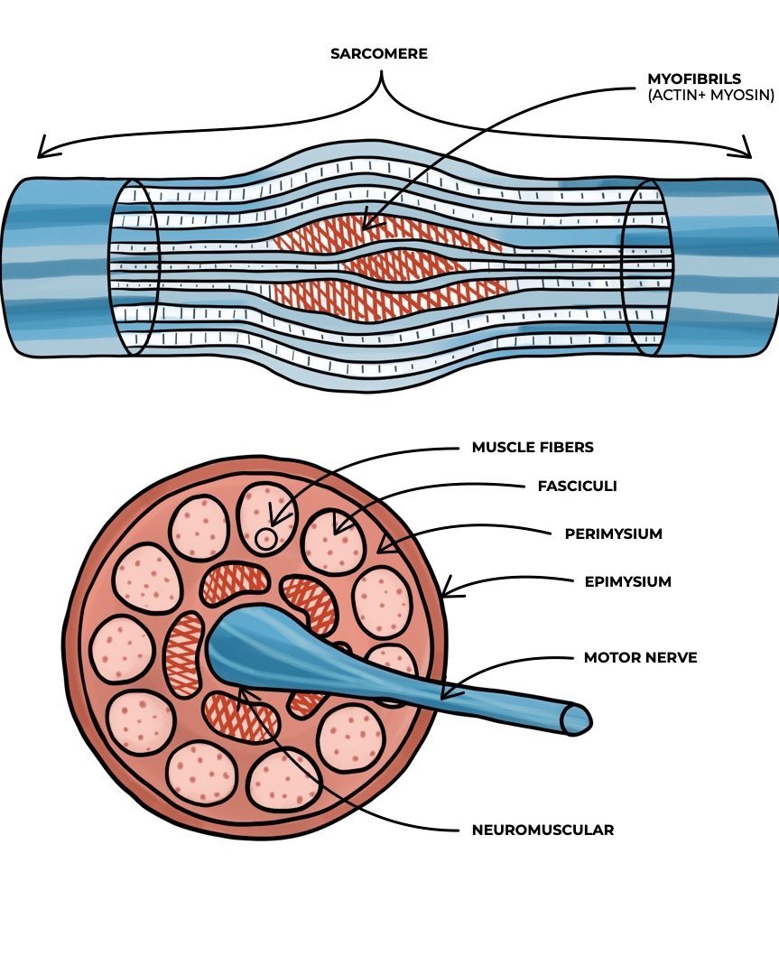
1.     Muscular Imbalances: Trigger points in muscles surrounding the SI joint, such as the piriformis, gluteus medius, or erector spinae muscles, can lead to muscular imbalances. When these muscles develop trigger points, they may become tight, weak, or painful, affecting the stability and function of the SI joint.

2.     Referral Pain: Trigger points can refer pain to other areas of the body, meaning that the pain is felt in a location distant from the trigger point itself. Trigger points in the muscles around the SI joint can refer pain to the lower back, buttocks, or even the hip, mimicking the symptoms of SI joint dysfunction.

3.     Altered Biomechanics: Muscular imbalances and trigger points can alter the biomechanics of the pelvis and lumbar spine, potentially placing additional stress on the SI joint. Over time, this altered biomechanics can contribute to SI joint dysfunction or exacerbate existing issues.

4.     Pain Amplification: Trigger points can sensitize the surrounding tissues and nervous system, making them more prone to pain and discomfort. This heightened sensitivity can make it more challenging for individuals with SI joint dysfunction to manage their pain effectively.

It's essential to note that trigger points are just one potential contributor to lower back and SI joint pain. SI joint dysfunction can have various causes, including trauma, inflammation, pregnancy-related changes, or degenerative processes. Addressing trigger points, if present, can be part of a comprehensive treatment plan to alleviate pain and improve function in individuals with SI joint issues.

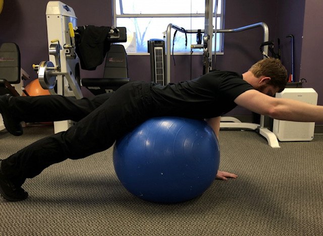
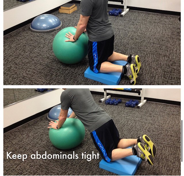
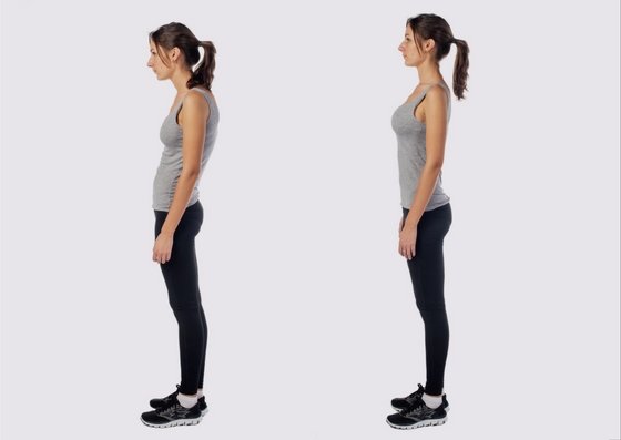
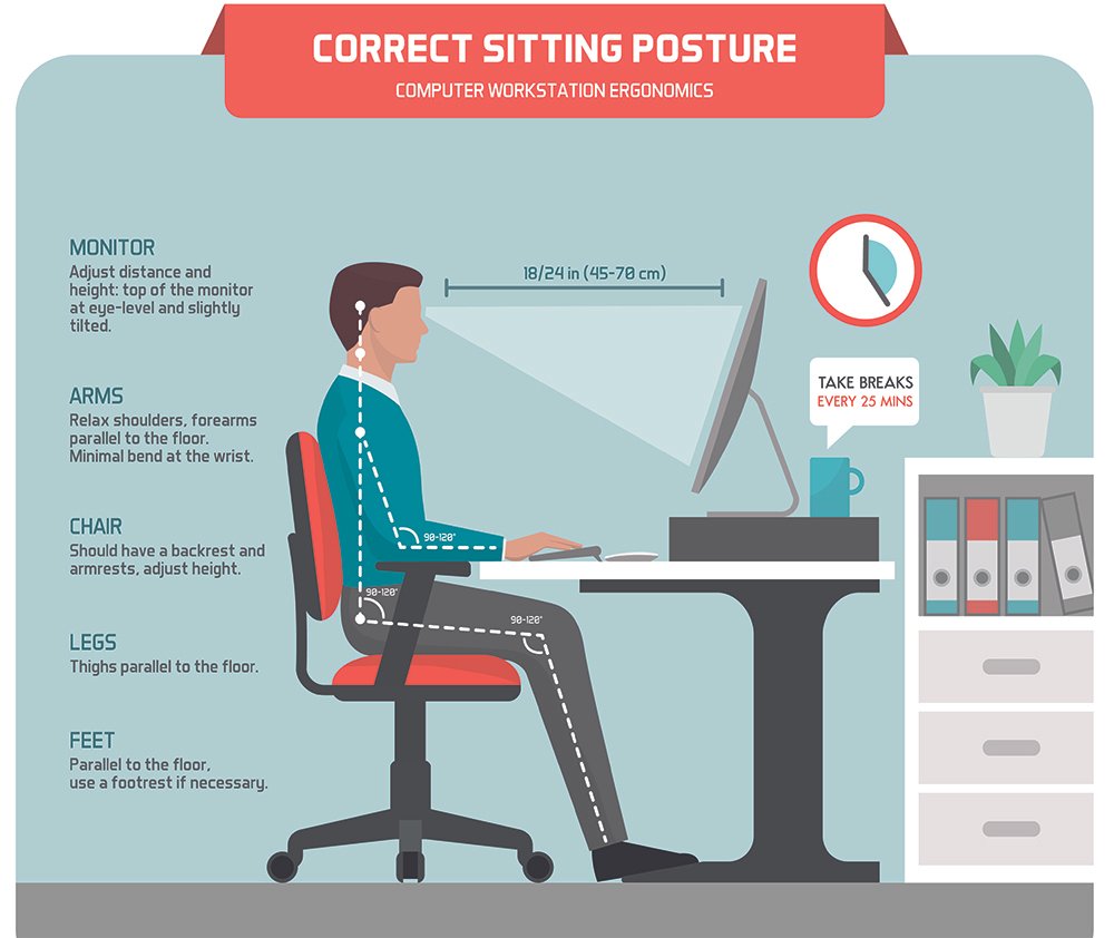
After a thorough examination, the physical therapist will discuss their findings and work with you to develop an individualized treatment plan. Treatment may include exercises to improve SI joint stability, manual therapy techniques, pain management strategies, and education on posture and body mechanics. The goal is to alleviate pain, improve function, and prevent future SI joint issues.

If you feel your pain may be related to a sacroiliac joint problem and would like to learn how to treat it successfully without surgery, please take advantage of a free consult at Lanier Therapy in Motion. 

Treating a Herniated Disc Non-Surgically

Let’s look at how trigger points can cause herniated discs in the lower back.  Bulging and herniated discs are relatively common.  It is estimated that if we were to MRI the lumbar spines of adults over the age of 30, 7 out of 10 would have bulging or herniated discs.  And 4 out of the 7 would still be asymptomatic and have no lower back pain.   The most common areas of the lumbar spine to have bulging or herniated discs are the L4-5 segment and the L5-S1 segment, in other words the last two lumbar segments where most spinal stress occurs. 

As the muscles become strained in this area, they shorten compressing the vertebra and cause the disc to bulge and eventually herniate due to the stress.  At this point, the nerve at this vertebral segment becomes inflamed producing back pain symptoms from active trigger points in the muscles surrounding the vertebra known as the deep paraspinals or Multifidus muscles.  The pain at this point will be located mostly in the lower back as illustrated below.

Multifidus Pain Pattern

Muscle Shortening caused by active trigger points will lead to compression of the discs and the facet joints leading to a bulging or herniated disc.  The disc will then begin to irritate the nerve at that segment.

Disc Bulge compressing a Lumbar Nerve

Once the nerve is irritated, active trigger points can form in the muscles innervated by that segment.  Let’s look at some of the typical trigger points caused by nerve compression at L4/5 and L5/S-1.  The L4/5 segment disc will typically begin to irritate the L5 nerve as it exits from the spinal column.  The L5 muscles which can be affected include the gluteal muscles, the hip rotators, and the hamstrings.  The L5/S1 segment disc will typically irritate the S1 nerve and will affect the S1 muscles such as the calves.  Typical trigger point referral patterns will be as follows:

L5 Gluteus Maximus Pain Pattern

L5 Gluteus Medius Pain Pattern

L5 Gluteus Minimus Pain Pattern

L5 Piriformis Pain Pattern

L5 Hamstrings Pain Pattern

S1 Gastrocnemius Pain Pattern

S1 Soleus Pain Pattern

Of course, these are not the only pain patterns caused by L4/5, and L5/S1 disc bulges and herniations.  Pain patterns vary and can also be accompanied by numbness, tingling and other nerve symptoms, including weakness in extreme cases.  In most cases, a physical therapist can assist you in developing a program to address the trigger points and reduce the disc herniation/ bulge.  There are exercise programs which are highly effective in treating trigger points and herniated discs.  In about 30% of cases, the affected segment may need special treatment such as trigger point dry needling to deactivate the trigger points enough to allow the disc to reduce and move back into place. 

Trigger Point Dry Needling the Multifidus Muscle

Trigger Point Dry Needling is a technique used to treat shortened muscles caused by trigger points. This type of therapy uses a “dry” non-cutting needle, without any injections or medications. The needle is inserted into the active trigger point which is causing the pain resulting in often immediate, dramatic, and lasting improvement.

This treatment is now under the scope of physical therapy and has improved the success rate in treating bulging and herniated discs.  The Physical Therapists at Lanier Therapy in Motion have extensive experience and have achieved dramatic results using Dry Needling to correct bulging and herniated discs. 


If you feel your pain may be related to a bulging or herniated disc and would like to learn how to treat it successfully without surgery, please take advantage of a free consult at Lanier Therapy in Motion. 

Back Trigger Points: Self-Treatment

BACK PAIN and TRIGGER POINTS

BACK PAIN SACROILIAC JOINT PAIN AND SCIATICA

Let’s look at the statistics of Lower Back Pain and its prevalence in the United States.

        1.      Prevalence: Low back pain and Sciatica (which is defined as pain in the lower extremity originating from a spinal source) is one of the most common reasons for seeking medical care in the U.S. It affects a large portion of the population at some point in their lives. Studies suggest that around 80% of adults will experience low back pain at some point.
        2.      Chronic Low Back Pain: While many cases of low back pain are acute and resolve within a few weeks, a substantial number of individuals develop chronic low back pain, which persists for three months or longer. Chronic low back pain can have a significant impact on daily functioning and quality of life. It is estimated that 20-30% of cases of lower back pain will become chronic.  Low Back pain recurrence is 90% with most recurrences happening every 18 months.  Up to 20-30% will become chronic requiring some type of surgical intervention.
        3.      Leading Cause of Disability: Low back pain is a leading cause of disability globally, and its impact is also notable in the U.S. It contributes to work absenteeism, decreased work productivity, and increased healthcare utilization.
        4.      Age Distribution: Low back pain is more common in adults, with the highest prevalence observed in those aged 30 to 60. Initial onset is usually at age 20-25.  However, it can affect individuals of all ages.
        5.      Gender: Studies suggest that women may be slightly more likely to experience low back pain compared to men.
        6.      Risk Factors: Risk factors for low back pain include factors such as sedentary lifestyles, obesity, smoking, poor posture, physically demanding occupations, and psychological factors like stress and depression.
        7.      Healthcare Utilization: Low back pain is a common reason for doctor visits, physical therapy appointments, and the use of pain medications. It contributes to a significant portion of healthcare costs.
        8.      Economic Impact: The economic burden of low back pain is substantial due to healthcare costs, lost work productivity, and disability. It is estimated to cost billions of dollars annually in the U.S

Common Lower Back Trigger Points

Quadratus Lumborum

Gluteus Maximus

Gluteus Medius

Gluteus Minimus

Piriformis

These trigger points can cause pain in the lower back, sacroiliac joint and down the leg in the case of sciatica.  If you feel your symptoms may be trigger point related, schedule a free consult with a Physical Therapist at Lanier Therapy in Motion for an evaluation and discussion of treatment options.

Neck Trigger Points: Self-Treatment

Once located, the trigger points can be best self-treated using a technique call ischemic compression massage or trigger point pressure release. 

TRIGGER POINT PRESSURE RELEASE

This technique involves the application of slowly increasing, nonpainful pressure over a trigger point until a barrier of tissue resistance is encountered.  Contact is then maintained until the tissue barrier releases, and pressure is increased to reach a new barrier to eliminate the trigger point tension and tenderness.  The pressure can be applied using a massage stick or cane, applying sustained pressure with a lacrosse ball or tennis ball, or by using a foam roller.  More recently, techniques to deactivate trigger points using percussion massage guns and vibrating foam rollers have become popular.  Let’s explore some of the techniques. 

TRIGGER POINT PRESSURE RELEASE METHODS


THERACANE

There are several versions of massage sticks or canes available.  The canes have several small balls along their length which allow precise application of pressure to the trigger points. These allow one to easily apply deep pressure massage on hard-to-reach areas such as the neck and back.  The technique involves applying adequate pressure to keep the ball in contact with the muscle group you’re working on and prevent the ball from slipping off. Begin with light pressure and short periods of use as your muscles will require a breaking in period.  Start off with just a minute or two of use a few times a day.  If you overdo it, you will likely hurt more than you did to begin with.  This discomfort will normally always go away after a restful sleep or within 12-36 hours.  In addition to this, a long hot shower or use of a heating pad will usually make you feel better and speed up your recovery.  With experience you’ll be able to use all the pressure you want for extended periods of massage. 

If a trigger point in the muscle seems extra sensitive, massage the surrounding area before working on the main trigger point. 

Try different methods when applying pressure to determine which suits you and/or the muscle you are working on. For example, you can apply steady pressure for 5 to 10 seconds, release, relax and repeat, or maintain direct pressure for 30 seconds to a minute.  Some prefer moving the ball slowly over a small area of the muscle, gradually going deeper.  A gentle wiggle can help you burrow into a sore area.

Lacrosse or Tennis Ball

A lacrosse ball or tennis ball can be used in a similar fashion to apply trigger point pressure release.  Place the ball at the site of the trigger point and apply pressure by lying on the ball.  If this is too painful, an alternate method would be to place the ball inside a sock to hold it in place and then use a wall to apply a gentler pressure.

Foam Roller

A less precise method for applying trigger point pressure (but still effective) is using a foam roller.  Lie on your back while placing the roller in the area of the trigger point.  Maintain the pressure for 30 seconds up to a minute or two before moving to another spot.  Begin with a few minutes of treatment and work up to 15 to 20 minutes once every day or two. 

Percussion Massage Gun

More recently, the percussion massage guns have been gaining popularity and for good reason.  They are highly effective in deactivating active trigger points in just a few sessions.  And typically the treatment is more relaxing and less painful than sustained pressure over the trigger point.  The massage ball on the percussion massage gun applies pressure to the trigger point in a rapid “on and off” method which prevents the typical painful perception when using sustained pressure over the trigger point.  The frequency of the percussion can be varied significantly for maximum comfort during the self-treatment session.  Massage guns can also be purchased that have less excursion or travel of the massage head for treating highly painful trigger points, while massage guns with maximum excursion can be used once the acute episode has subsided and the trigger point is less painful. 

Because of this, percussion massage guns are now my recommended method for self-treatment of trigger points.  The massage guns are very affordable and highly effective. 

Vibrating Foam Roller

Similar to the percussion massage gun, the vibrating foam roller applies an “on and off” pressure to the trigger point.  The frequency of the vibration can be varied and the amplitude of the vibration is much smaller than that of the percussion massage gun.  For this reason, they are recommended for initial treatment of highly sensitive trigger points or trigger points close to the spine.  The treatment technique is the same as for a regular foam roller.

Physical Therapists are highly skilled in using your symptoms and palpation techniques to isolate trigger points.  If you feel your symptoms may be trigger point related, schedule a free consult with a Physical Therapist at Lanier Therapy in Motion for an evaluation and discussion of custom self-treatment options.

Neck Pain and Trigger Points

Neck pain is a common medical condition that can have a significant impact on an individual's quality of life. Understanding its epidemiology involves looking at its prevalence, risk factors, and associated factors.

1.    Prevalence: Neck pain is a widespread issue globally. Its prevalence varies by age, gender, and geographic location. Generally, it is more prevalent in adults than in children and adolescents. Studies have shown that the lifetime prevalence of neck pain is up to 70%, while the annual prevalence can vary from 10% to 20%.

2.    Gender Differences: Neck pain appears to be more common in women than in men, although the reasons for this are not entirely clear. Hormonal differences, differences in anatomy, and variations in pain perception may play a role.

3.    Age: Neck pain is most commonly reported by individuals between the ages of 30 and 60. However, it can affect people of all age groups.

4.    Occupation: Occupational factors can significantly contribute to neck pain. Jobs that involve prolonged sitting, repetitive neck movements, or poor ergonomics are associated with a higher risk of developing neck pain. This is especially relevant in office workers, healthcare professionals, and those in physically demanding professions.

5.    Psychosocial Factors: Stress, anxiety, and depression can contribute to the development and persistence of neck pain. Individuals with high levels of stress or poor mental health are more likely to report neck pain.

6.    Lifestyle Factors: Sedentary lifestyles, lack of physical activity, and poor posture are risk factors for neck pain. Modern technology, such as extended use of computers and mobile devices, has also been linked to an increase in neck pain cases.

7.    Trauma and Injury: Acute neck pain can result from trauma or injury, such as whiplash from car accidents, sports injuries, or falls.

8.    Chronic Conditions: Certain chronic medical conditions like osteoarthritis, degenerative disc disease, and herniated discs can lead to chronic neck pain.

The percentage of neck problems that eventually become chronic can vary depending on the specific population, the definition of chronic neck problems, and the study methodology. However, chronic neck pain is generally considered when the condition persists for three months or longer. Here are some approximate figures:

1.    Overall Chronicity Rate: Research suggests that around 10-20% of cases of acute neck pain progress to become chronic neck pain.

2.    Recurrent Neck Pain: It's important to note that 50-85% of cases can be episodic or recurrent, meaning it comes and goes over time. Recurrent neck pain can also impact an individual's quality of life, even if it doesn't remain chronic.

3.    Chronic Neck Pain Risk Factors: Certain factors can increase the likelihood of acute neck pain becoming chronic. These include psychological factors (such as anxiety and depression), high levels of pain-related disability at the outset, and poor response to initial treatments.

4.     Specific Conditions: Some underlying conditions, such as degenerative disc disease or cervical radiculopathy, may have a higher likelihood of leading to chronic neck pain if left untreated or if not managed appropriately. DDD is a condition characterized by the gradual degeneration of the intervertebral discs in the cervical spine. It can lead to chronic neck pain, as well as other symptoms like radiating arm pain, numbness, and weakness. The percentage of individuals with DDD who develop chronic neck pain is not precisely defined, but it's estimated that a significant portion of people with advanced DDD will experience persistent or recurrent neck pain.

5.    Occupational Factors: Chronicity rates may be higher in individuals whose neck pain is related to their occupation, especially if the job involves repetitive motions or poor ergonomics.

It's important to remember that chronic neck pain can have a significant impact on an individual's daily life, emotional well-being, and work productivity. Early intervention, proper diagnosis, and a tailored treatment addressing active trigger points can help prevent or manage chronic neck pain effectively.

Common Neck Trigger Points

In order to treat your neck and upper back trigger points, first we must identify them.  Usually you can find them because they feel like “knots” or “tight bands” within the muscle and will reproduce your symptoms when you press on them.  You may also be able to find them by looking at typical trigger point pain referral patterns.  Here are some of the more common ones. 

Trapezius Trigger Points

Levator Scapula Trigger Points

Rhomboid Trigger Points

Serratus Posterior Superior Trigger Points

Scalene Trigger Points

Physical Therapists are highly skilled in using your symptoms and palpation techniques to isolate trigger points.  If you feel your symptoms may be trigger point related, schedule a free consult with a Physical Therapist at Lanier Therapy in Motion for an evaluation and discussion of treatment options.

More about Trigger Points and Myofascial Pain

Here are some key points to know about trigger points:

1.    Causes: Trigger points can be caused by a variety of factors, including muscle overuse, poor posture, muscle imbalances, direct trauma, stress, and even chronic medical conditions. Repetitive motions, such as typing or lifting heavy objects, can contribute to the development of trigger points.

2.    Symptoms: Trigger points often cause local pain or discomfort in the affected muscle. They can also lead to referred pain, where pain is felt in other areas of the body. For example, a trigger point in the neck or shoulder could cause pain that radiates down the arm. Other symptoms might include muscle stiffness, limited range of motion, and muscle weakness.

3.    Palpation: Healthcare professionals, such as physical therapists or massage therapists, can often locate trigger points by palpating the muscle tissue. Trigger points can feel like small nodules or knots within the muscle. When pressure is applied to a trigger point, it can often reproduce the pain or discomfort that a person is experiencing.

4.    Treatment: There are several approaches to treating trigger points, depending on their severity and underlying causes. Some common treatments include:

  • o   Manual Therapy: Massage therapy, myofascial release, and other hands-on techniques can help release tension in the muscle and alleviate trigger point-related pain.

  • o   Stretching and Exercise: Gentle stretching and specific exercises can help relax the muscle and prevent the formation of new trigger points. Strengthening weak muscles can also help improve muscle imbalances that might contribute to trigger points.

  • o   Heat and Cold Therapy: Applying heat or cold to the affected area can help relax the muscle and reduce pain. Heat can help increase blood flow, while cold can help reduce inflammation.

  • o   Trigger Point Injections: In some cases, a healthcare provider might inject a local anesthetic or other medication directly into the trigger point to alleviate pain and reduce muscle tension.

  • o   Medications: Over-the-counter pain relievers, such as nonsteroidal anti-inflammatory drugs (NSAIDs), can help manage pain and inflammation associated with trigger points.

5.    Prevention: To prevent trigger points from forming or recurring, it's important to maintain good posture, practice regular stretching and exercise, stay hydrated, manage stress and avoid prolonged periods of overuse.

Trigger points are thought to be areas of hyperirritable muscle tissue that are characterized by tight bands or knots. They can develop in both skeletal and smooth muscles. The exact mechanisms behind trigger point formation and their effects are not fully understood, but there are a few theories that help explain what might be happening within the muscle:

1.    Microtrauma and Muscle Fiber Dysfunction: One theory suggests that trigger points can form due to microtrauma or small injuries to muscle fibers. These injuries can be caused by overuse, strain, or direct trauma. When muscle fibers become damaged, they can contract and tighten, forming taut bands within the muscle. This contraction can restrict blood flow to the affected area, leading to a decrease in oxygen and nutrients, and an accumulation of waste products.

2.    Neuromuscular Junction Dysfunction: Another theory proposes that trigger points may result from dysfunction at the neuromuscular junction—the point where nerves communicate with muscle fibers. It's believed that excessive nerve impulses can cause muscle fibers to contract and remain in a shortened state, leading to the formation of trigger points.  Typically the nerve is in an irritated state due to compression in or around the spine or along its course through the myofascial chain.

3.    Energy Crisis Hypothesis: This theory suggests that trigger points develop due to an energy crisis within muscle cells. When muscle fibers are overused or stressed, they can become depleted of energy-rich molecules like ATP. This energy deficit can lead to a sustained state of contraction in localized muscle fibers, contributing to the development of trigger points.

4.    Motor Endplate Hypothesis: According to this theory, trigger points form due to dysfunctional motor endplates, which are specialized areas where nerve terminals connect with muscle fibers. The communication between nerves and muscle fibers can become disrupted, leading to muscle fiber contraction, reduced blood flow, and the formation of trigger points.

5.    In addition to the underlying mechanisms, trigger points can cause a cycle of pain and dysfunction. When a trigger point forms, it can cause the muscle to become shortened and tense, which can lead to decreased blood circulation, oxygen deprivation, and a buildup of metabolic waste products like lactic acid, substance P, CGRP, prostaglandins and bradykinins.  This can further irritate nerve endings and contribute to pain. The pain itself can lead to protective muscle guarding, which can perpetuate the cycle by causing additional muscle tension and trigger point formation.

6.    It's important to note that the science behind trigger points is still evolving, and there is ongoing research to better understand their causes and mechanisms. Treatment approaches are often based on relieving muscle tension, promoting blood flow, and addressing any underlying factors that contribute to their development.

Let’s take a look at the anatomy of the muscle fiber itself to see what is happening to cause a trigger point: Notice in the diagram that the myofibrils within the sarcomere are in a semicontracted state due to a local dysfunction.  The cause of this dysfunction can vary. 

There are a few theories to explain why the trigger point is not always located in the painful area.   Referred pain is a phenomenon in which pain is perceived in an area of the body that is remote from the actual source of the pain. This can often be confusing and challenging to diagnose because the pain appears to be coming from a location that is not directly related to the underlying issue. Referred pain occurs due to the complex network of sensory nerves and the way the brain interprets these signals. Here are a few of the proposed theories.

1.    Nerve Convergence: Nerves from different parts of the body often converge or come together and share pathways as they travel to the spinal cord and brain. This convergence can lead to a phenomenon known as "cross-talk" between nerve pathways. When a trigger point is active and sending pain signals to the spinal cord, the brain might interpret those signals as originating from a different area of the body that shares nerve pathways with the affected muscle.

2.    Central Sensitization: Trigger points can sensitize the central nervous system, which includes the brain and spinal cord. This means that the nervous system becomes more sensitive to pain signals. When a trigger point is active, it can heighten the sensitivity of nerves in the spinal cord, amplifying pain signals. As a result, the brain might perceive pain as coming from a larger area than just the trigger point itself.

3.    Dermatomes and Innervation Patterns: Dermatomes are specific regions of the skin that are innervated by a single spinal nerve. Each spinal nerve carries sensory information from a particular area of the body. When there is convergence of nerve pathways in the spinal cord, pain signals from a specific organ or muscle might be perceived in the dermatome associated with the same spinal nerve.


4.    Referred Pain Patterns: Some trigger points have consistent patterns of referred pain that are well-documented. These patterns are thought to be related to the specific muscle's innervation and nerve connections. For example, a trigger point in the trapezius muscle in the upper back might refer pain down the arm and into the hand, into the side of the scalp or the lower jaw, even though the trigger point is located in the upper back.

5.    Neural Sensitization: Trigger points can cause abnormal sensory signals to be sent to the brain. These abnormal signals can lead to the brain interpreting the sensations as pain in different areas of the body. Additionally, the nervous system's response to the presence of a trigger point can cause surrounding muscles to tense up and contribute to the spread of pain.

Referred pain can sometimes make diagnosing the source of pain challenging, as the actual cause of the pain may be located quite far from where the pain is felt. Healthcare professionals, such as physical therapists and doctors, often rely on their knowledge of anatomy, pain patterns, and patient history to help determine the true source of the pain. Usually, a thorough evaluation to identify the source of the pain and develop an appropriate treatment plan.


Trigger points can be present along the course of a dermatome or myotome. 

Dermatomes and myotomes are an important concept in understanding how referred pain from trigger points occurs. Let's explore in more detail:

1.    Dermatomes:

A dermatome is a specific region of the skin that is innervated by sensory nerve fibers from a single spinal nerve. These nerve fibers transmit sensory information such as touch, temperature, and pain from the skin to the spinal cord and then to the brain. Each spinal nerve is responsible for supplying sensory input to a specific dermatome.

2.    Dermatomes are arranged in a segmented pattern along the body, with overlapping regions. Because of this overlap, adjacent dermatomes share some degree of sensory innervation. This overlap plays a crucial role in the phenomenon of referred pain.

3.    A myotome refers to a group of muscles that are innervated by the same segment of the spinal cord. In other words, a myotome is a region of the body that is primarily controlled by a single spinal nerve or a specific segment of the spinal cord. Myotomes play a critical role in muscle movement and coordination throughout the body.  Trigger points may be located in muscles innervated by a specific spinal segment.

4.    Each spinal nerve in the human body is associated with a specific myotome, and these myotomes are responsible for providing motor control to various muscle groups. When a motor nerve signal is sent from the spinal cord to a particular myotome, it triggers the contraction of the muscles within that myotome, leading to coordinated movement.  Muscles fibers that contain trigger points have been shown to be twice under twice as much resting tension, and twice as weak as a regular muscle fiber. This affects all coordinated movement along the myotome.

5.    The concept of myotomes is often used in medical and clinical contexts, such as in the assessment of muscle strength and the diagnosis of neurological conditions. Healthcare professionals may evaluate specific myotomes to determine the extent of muscle weakness or dysfunction associated with trigger points.

6.    Referred pain occurs when the brain misinterprets sensory signals and perceives pain in an area of the body different from the actual source of the pain. This often happens because the pain signals from an internal organ or deep tissue converge onto the same spinal nerve pathways that supply the skin in a certain dermatome or myotome.

7.    For example, let's consider the classic example of a heart attack. The heart is supplied by sensory nerves that converge onto spinal nerves in the upper thoracic region of the spinal cord. These spinal nerves also supply the skin over the chest and upper left arm. As a result, during a heart attack, the brain might interpret the intense pain from the heart as originating from the chest and left arm—the same dermatome that shares nerve pathways with the heart.

8.    In cases like this, the pain is referred to the skin because the sensory nerve pathways from the affected organ and the skin overlap in the spinal cord. This creates a connection between the internal source of pain and the skin in a specific dermatome

9.    Understanding dermatomes/myotomes and referred pain is important in medical diagnosis. When a patient reports pain in a certain area, healthcare professionals can use their knowledge of dermatomes and myotomes to locate the active, latent, and satellite trigger points.

10. However, it's important to note that while referral patterns are generally consistent, individual variation can occur. Some people might experience slightly different referred pain patterns due to their unique nerve connections.

Typical pain referral pattern for a trigger point in the Serratus Posterior Superior muscle.

If you feel your pain could be trigger point related, schedule now for a free consult with a physical therapist at Lanier Therapy in Motion to look at possible treatment options.

Myofascial Pain and Trigger Points

WHAT ARE “TRIGGER POINTS”?


Skeletal muscle is the largest single organ in the human body and accounts for nearly 50% of body weight.  There are over 650 named muscles in the human body.  Any one of these muscles can develop myofascial trigger points that refer pain and motor dysfunction, often to another location.  Yet the muscles in general and trigger points in particular receive little attention as a major source of pain and dysfunction in modern medical school teaching and medical textbooks.  In addition, trigger points do not show up in x-rays, MRI’s or other routine tests.  The only way to identify them is with hands on manual palpation, of which today’s doctors have precious little time to perform. 


Trigger points were first identified and described by Dr. Janet G. Travell and Dr. David G. Simons. They were pioneering physicians in the field of pain management and musculoskeletal medicine. Their work on trigger points began in the mid-20th century, and they published their findings in their influential two-volume book “Myofascial Pain and Dysfunction: The Trigger Point Manual,” which was first published in 1983.

As the story goes, Janet Travell was doing rounds in a cardiac ward, many of the patients would complain of pain in the chest wall, thoracic or scapular regions.  The cardiologists would tell the patients: “Of course you’re hurting… you just had heart surgery.”  Later, Travell was in a pulmonary ward.  The patients would complain of the same type of pains.  The pulmonologists would respond: “Of course you have pain there, you just had lung surgery.”  And later as she was doing rounds in a general ward when the patients would complain, the general practitioners would respond with: “It’s psychosomatic!”  But Travell began to notice that during manual examination of these patients she could locate palpable tight bands within the chest wall, cervical and thoracic musculature that would reproduce the patient’s pain complaints.  The patients would respond with “You found it!” 

Dr. Travell was a White House physician during the Kennedy and Johnson administrations, and her observations of musculoskeletal pain patterns in her patients led to the development of the concept of trigger points. Dr. Simons, a neurologist, collaborated with Dr. Travell to further investigate and document trigger points.

Their research involved extensive clinical observations, manual examination of patients, and the use of injection techniques to identify and treat trigger points. Through their work, they mapped out the locations of trigger points in various muscles, documented the referred pain patterns associated with them, and developed techniques for their treatment.

Today, trigger point therapy is a widely recognized and utilized approach in pain management and physical therapy, thanks to the pioneering work of Dr. Travell and Dr. Simons.

Trigger points, also known as muscle knots, are sensitive areas within the muscle fibers that can cause pain and discomfort. These points are usually found in taut bands of muscle and are often associated with muscle tension, overuse, or injury. Trigger points can occur in various muscles throughout the body and can cause referred pain, which means that the pain is felt in a different area of the body than where the trigger point is located.

Notice how a trigger point in the gluteal musculature can refer pain all the way to the calf and ankle.

 TYPES OF TRIGGER POINTS

Trigger points have been categorized into three main types: active, latent, and satellite trigger points.  The main difference between them lies in their level of symptom presentation and their influence on pain and discomfort:


  1. Active Trigger Points:

    • Symptoms: Active trigger points are characterized by causing spontaneous pain or discomfort, even at rest. They can produce a constant or intermittent dull ache or sharp pain, which may be localized to the trigger point itself or can refer pain to other areas of the body.

    • Sensitivity: Active trigger points are tender to the touch. Palpation of these points often reproduces the pain that the person is experiencing, and pressing on them might cause a twitch response in the muscle.

    • Influence: Active trigger points are the primary source of pain and dysfunction. They can significantly affect muscle function, range of motion, and overall quality of life.

  1. Latent Trigger Points:

    • Symptoms: Latent trigger points are dormant and do not actively produce pain or discomfort unless they are pressed or stimulated. They might not cause noticeable symptoms on their own.

    • Sensitivity: Latent trigger points can still be tender when pressure is applied, but they do not generate the same level of spontaneous pain as active trigger points.

    • Influence: Latent trigger points can contribute to muscle imbalances and restricted movement. They may become active trigger points if they are subjected to additional stress or strain.

  1. Satellite Trigger Points:

    • Definition: Satellite trigger points are secondary points that can develop in response to an existing active trigger point. They are located in muscles that are functionally related or anatomically connected to the muscle with the active trigger point.

    • Symptoms: Satellite trigger points can refer pain and discomfort similar to active trigger points. However, their pain is often felt in areas away from the original active trigger point.

    • Influence: Satellite trigger points can complicate the pain pattern and contribute to the spread of discomfort. Treating both the primary active trigger point and any satellite trigger points is important for comprehensive pain management.


Active trigger points are the primary culprits for causing pain and discomfort, while latent trigger points are inactive until triggered by pressure or other stimuli. Satellite trigger points are secondary points that develop as a result of the presence of an active trigger point, and they can refer pain to different areas. All three types of trigger points—active, latent, and satellite—can play a role in muscle pain and dysfunction, and addressing them as part of a comprehensive treatment plan can help alleviate symptoms and improve muscle function.

What is the difference between “Trigger Points” and “Tender Points?”


The main difference between trigger points and tender points lies in their characteristics and associated conditions:  Trigger points are localized knots or tight bands of muscle fibers that can cause referred pain when pressed or stimulated. They are typically associated with myofascial pain syndrome, which is a condition characterized by muscle pain and discomfort. Trigger points often have specific referral patterns, meaning they can cause pain or discomfort in areas distant from the actual point.

Tender points are specific anatomical locations on the body that are particularly sensitive to touch or pressure. They are not abnormal although they may become particularly sensitive as in the case of fibromyalgia.  They are a defining feature of fibromyalgia, a chronic pain disorder. To be diagnosed with fibromyalgia, a person must have a certain number of tender points. Tender points are more diffuse and widespread than trigger points and are not associated with referred pain patterns.

In summary, trigger points are localized abnormal muscle knots that can cause referred pain, often associated with myofascial pain syndrome. Tender points, on the other hand, are specific, sensitive points on the body associated with fibromyalgia and are not typically associated with referred pain.

TREATMENT


Physical therapy can be an effective approach to treating trigger points and addressing the pain and discomfort they cause. Physical therapists use a variety of techniques to relieve muscle tension, improve muscle function, and reduce pain. Here are some common physical therapy techniques used to treat trigger points:

  1. Manual Therapy:

    • Massage: Therapists use various massage techniques to manipulate and knead the affected muscles. This can help relax muscle fibers, improve blood circulation, and reduce muscle tension.

    • Myofascial Release: This technique focuses on releasing tension in the fascia, the connective tissue that surrounds muscles. Myofascial release involves gentle stretching and sustained pressure to alleviate muscle tightness.

    • Trigger Point Release: Therapists apply direct pressure to the trigger point itself to release tension and reduce pain. This can be done using their fingers, thumbs, or specialized tools.


2.    Trigger point dry needling:

Trigger point dry needling is a technique used in physical therapy to treat trigger points and alleviate muscle pain and tension. It involves the insertion of thin, solid needles into the skin and muscle directly at the site of the trigger point. The goal of trigger point dry needling is to release muscle tension, improve blood flow, and promote healing.


  1. Stretching Exercises:

    • Static Stretching: Targeted stretches are used to elongate and relax muscles, which can help alleviate muscle tension and reduce the impact of trigger points.

    • Dynamic Stretching: Gentle, controlled movements are used to gradually increase the range of motion and flexibility of the affected muscles.

4.    Strengthening Exercises:

·       Muscle Imbalance Correction: Strengthening exercises focus on addressing muscle imbalances that might contribute to the formation of trigger points. Strengthening weak muscles can help improve overall muscle function and reduce strain on other muscles.

·       Postural Correction: Therapists prescribe exercises to improve posture, as poor posture can lead to muscle imbalances and increased tension.

5.    Heat and Cold Therapy:

    • Heat Application: Heat can help relax muscles, increase blood flow, and promote healing. Therapists might use heat packs or hot towels on the affected area.

    • Cold Application: Cold therapy can reduce inflammation and numb the area, helping to alleviate pain. Cold packs or ice massages might be used.

  1. Ultrasound and Electrical Stimulation:

    • Ultrasound: Therapists use ultra high frequency sound waves to deliver deep heat to the affected tissues. This can increase blood flow and promote healing.

    • Electrical Stimulation: Electrical currents can be applied to the muscles to reduce pain and relax muscle fibers.

  1. Home Exercises and Self-Care:

    • Patients are often given specific exercises and stretches to continue at home, along with self-massage techniques and strategies for managing pain.

  1. Posture and Body Mechanics Education:

    • Therapists provide guidance on maintaining proper posture and body mechanics during daily activities to prevent further trigger point development.


It's important to note that the treatment approach may vary based on the individual's condition, the severity of the trigger points, and any underlying medical issues. Physical therapists will conduct a thorough evaluation to determine the most appropriate treatment plan.

The goal of physical therapy for trigger points is to reduce pain, improve muscle function, and prevent the recurrence of trigger points. Patients are often advised to maintain an active lifestyle, practice good posture, manage stress, and perform their prescribed exercises consistently to achieve the best results.


If you feel your pain could be trigger point related, schedule now for a free consult with a physical therapist at Lanier Therapy in Motion to look at possible treatment options.

Align Your Spine.... Final Tips

Our summary video available on our YouTube channel @LanierTherapyinMotion on learning how to align your spine to treat and prevent neck and back pain.

In addition to strengthening the core, strengthening the muscles used to perform heavy lifting may also be beneficial with long term management of lower back trigger points.  Some good prevention exercises would include the following:

Squats:

    • Targets: Quadriceps, hamstrings, glutes, lower back, core.

Deadlifts:

    • Targets: Hamstrings, glutes, lower back, upper back, core.

Lunges:

    • Targets: Quadriceps, hamstrings, glutes, core.

Planks:

    • Targets: Core, shoulders, lower back.

Bent-Over Rows:

    • Targets: Upper back, lats, biceps.

Hip Thrusts:

    • Targets: Glutes, hamstrings, lower back, core.

Russian Twists:

    • Targets: Obliques, core.

Farmers Walk:

    • Targets: Grip strength, forearms, core.

Remember to start with weights that are manageable and gradually increase the weight as you build strength. Always prioritize proper form over the amount of weight lifted.


Proper sleeping positions are also important for long term management of lower back and neck trigger points.  Night time may be particularly problematic with trigger points because the muscle is in a constant state of contraction which compresses nerves and blood vessels. During the day while we are changing positions and moving, the tissue continues to receive nutrition, but at night when movement is eliminated pain chemicals can build up in the affected muscles resulting in increased pain at night or first thing in the morning. 

Back Sleeping (Supine Position):

    • Pillow Support: Use a supportive pillow that keeps your head aligned with your spine. Consider using a thin pillow under your knees to maintain the natural curve of your lower back.

    • Mattress Support: Choose a medium-firm mattress that provides adequate support for your spine while contouring to your body's curves.

    • Avoid Arms Underneath: Avoid sleeping with your arms positioned under your head or pillow, as this can lead to shoulder strain.

    • Neutral Alignment: Back sleeping tends to maintain a more neutral alignment for your spine, reducing the risk of unnecessary twisting or pressure on certain areas.

Side Sleeping (Sidelying):

    • Pillow Support: Place a pillow between your knees to keep your hips aligned and prevent your top leg from pulling your spine out of alignment. Use a supportive pillow for your head that keeps your neck in line with your spine.

    • Mattress Support: Similar to back sleeping, choose a mattress that provides proper support and comfort for your body.

    • Curled Fetal Position: Sleeping with your knees drawn up toward your chest can help relieve pressure on your lower back. However, make sure not to curl up too tightly, as this can lead to strain over time.

    • Alternate Sides: If you're a side sleeper, try alternating sides to avoid overloading one side of your body consistently.

Stomach Sleeping (Prone Position):

    • Consider Alternatives: Sleeping on your stomach is generally not recommended, as it can strain your neck and lower back. If possible, try transitioning to back or side sleeping.

    • Pillow Placement: If you must sleep on your stomach, use a thin pillow or no pillow at all for your head. This helps reduce strain on your neck.

    • Flat Surface: Choose a firm mattress to provide adequate support for your spine and minimize sinkage.

Remember that individual preferences and body types can influence which sleeping position is most comfortable for you. While these guidelines can help, it's important to prioritize your comfort and ensure that your chosen sleeping position supports your spine's natural curves.


When it comes to choosing a good mattress brand, there are several reputable options available. Keep in mind that mattress preferences can be quite subjective, so it's a good idea to test out mattresses in person if possible before deciding. Here are some well-known mattress brands that are generally recognized for their quality:

1.     Tempur-Pedic: Tempur-Pedic is known for its memory foam mattresses that offer excellent support and pressure relief. They are often praised for their durability and ability to conform to the body's shape.

2.     Saatva: Saatva offers a variety of luxury mattresses, including innerspring, memory foam, and hybrid options. Their mattresses are known for their quality materials and craftsmanship.

3.     Casper: Casper is a popular online mattress brand that offers a range of memory foam and hybrid mattresses. They are known for their comfortable designs and customer-friendly policies.

4.     Purple: Purple mattresses are unique for their hyper-elastic polymer comfort layer, which provides good support and breathability. They're especially well-regarded for their ability to alleviate pressure points.

5.     Sealy: Sealy has been a trusted name in mattresses for decades. They offer a wide range of options, including innerspring, memory foam, and hybrid mattresses.

6.     Beautyrest: Beautyrest mattresses are known for their pocketed coil technology that provides excellent support and motion isolation. They offer a variety of comfort levels and styles.

7.     Tuft & Needle: Tuft & Needle specializes in simple, budget-friendly foam mattresses that provide good support and comfort.

8.     Helix: Helix offers personalized mattresses based on a sleep quiz that assesses your preferences and needs. This customization can help you find a mattress that's tailored to you.

9.     Nectar: Nectar mattresses are known for their affordability and pressure-relieving memory foam layers.

10. Avocado: Avocado specializes in eco-friendly and organic mattresses made from natural materials. They offer latex and hybrid options.

When choosing a mattress, consider factors such as your preferred sleeping position, firmness preferences, any specific needs you may have (e.g., back pain, allergies), and your budget. Reading customer reviews and doing thorough research can also help you make an informed decision. If possible, visit mattress showrooms to test out different options and find the one that provides the right combination of comfort and support for you.


Side sleepers often benefit from mattresses that provide adequate pressure relief for the hips and shoulders, as well as proper spinal alignment. Here are some mattress types that are generally well-suited for side sleepers:

1.     Memory Foam Mattresses: Memory foam mattresses contour to the body's shape, providing excellent pressure relief for the hips and shoulders. They can help alleviate pain and discomfort that side sleepers might experience.

2.     Hybrid Mattresses: Hybrid mattresses combine the support of innerspring coils with the comfort of memory foam or latex. They often offer a good balance of support and pressure relief for side sleepers.

3.     Latex Mattresses: Latex mattresses are known for their responsiveness and support. They can provide the cushioning needed for the hips and shoulders while maintaining proper spinal alignment.

4.     Soft to Medium Firmness: Side sleepers generally benefit from mattresses that fall in the soft to medium firmness range. These firmness levels help contour to the body's curves and prevent pressure points.

5.     Zoned Support Mattresses: Some mattresses have zoned support, meaning they are designed with different firmness levels in different areas. Zoned support can provide extra cushioning for the shoulders and hips while maintaining support for the rest of the body.

6.     Pillow-top or Euro-top Mattresses: These mattresses have an additional layer of cushioning sewn onto the top, providing extra comfort and pressure relief for side sleepers.


Remember that mattress preferences can be subjective, so it's essential to consider your own comfort and needs. If possible, try out mattresses in person to see which one feels the most comfortable for your side-sleeping position. Also, pay attention to factors such as your body weight, as heavier individuals might require slightly firmer mattresses for proper support.


Back sleepers generally benefit from mattresses that provide proper support to the natural curves of the spine while maintaining a neutral alignment. Here are some mattress types that are often well-suited for back sleepers:

1.     Medium Firm to Firm Mattresses: Back sleepers typically require a mattress with medium firmness to firmness. This helps keep the spine properly aligned and prevents sinking too deeply into the mattress.

2.     Memory Foam Mattresses: Memory foam mattresses can contour to the body's shape, providing support to the lower back and maintaining proper alignment.

3.     Latex Mattresses: Latex mattresses offer responsiveness and support. They can help keep the spine aligned while providing comfort and cushioning.

4.     Hybrid Mattresses: Hybrid mattresses with a combination of innerspring coils and foam or latex layers can offer both support and pressure relief for back sleepers.

5.     Zoned Support Mattresses: Mattresses with zoned support can provide targeted support to different areas of the body, ensuring proper spinal alignment for back sleepers.

6.     Adjustable Mattresses: Some mattresses allow you to adjust the firmness levels on each side of the bed, which can be particularly useful if you share the bed with a partner who prefers a different sleep position.

7.     Innerspring Mattresses with Pillow-top: Innerspring mattresses with a pillow-top or euro-top layer can provide extra cushioning and support for back sleepers.

8.     Temperature-Regulating Mattresses: Back sleepers who tend to sleep hot might benefit from mattresses with cooling technologies, such as gel-infused foam or breathable materials.

Ultimately, the best mattress for you as a back sleeper depends on your personal preferences and comfort level. If possible, try out mattresses in stores to find the one that feels most supportive and comfortable for your back-sleeping position.


In addition to proper mattress selection, orthopedic pillows and supports can be incredibly beneficial for maintaining proper spinal alignment and reducing discomfort, especially during sleep. Here are some recommendations for orthopedic pillows and supports that can help improve your sleep quality and overall spinal health:

Orthopedic Pillows:

    • Contour Pillow: Contour pillows have a curved design that supports the natural alignment of your head, neck, and shoulders. They can help alleviate neck pain and reduce pressure points.

    • Cervical Pillow: Cervical pillows are designed to provide support to the cervical spine (neck). They often have a higher area to support the neck curve and a lower area to support the head.

    • Memory Foam Pillow: Memory foam pillows conform to the shape of your head and neck, providing customized support and pressure relief.

    • Wedge Pillow: A wedge pillow can be placed under your upper body to elevate the upper torso slightly. This can be helpful for conditions like acid reflux or snoring, while also providing support to the spine.

Orthopedic Supports:

    • Lumbar Support Cushions: Lumbar support cushions, called night rolls or sleep rolls are designed to provide night time support to the lower back's natural curve.

    • Knee Pillow: Placing a knee pillow between your knees while sleeping on your side can help maintain proper alignment of your hips and reduce strain on your lower back.

Full Body Pillows:

    • Body Pillows: Full body pillows can provide support to your entire body, particularly during side sleeping. They help align the spine, support the hips, and reduce strain on the neck and back.

Remember that the effectiveness of these products can vary based on individual needs and preferences.  

MEDICAL DISCLAIMER

Although we are physical therapists by profession, we are not YOUR physical therapist.  All content and information in this blog is for informational and educational purposes only, does not constitute medical advice, and does not establish any kind of patient-client relationship by your use of this blog.   A patient-client relationship with you is only formed after we have expressly entered into a written agreement with you that you have signed including our fee structure and other terms to represent you in a specific matter.  Although we strive to provide accurate general information, the information presented here is not a substitute for any king of professional advice, and you should not rely solely on this information.  Always consult a professional in the area for your particular needs and circumstances prior to making any professional, legal, medical and financial or tax-related decisions.

Best exercises for maintaining spinal alignment

A Closer look at the best exercises to maintain your spinal alignment falls into two categories.

1.     Exercises that promote the core stability required to maintain proper pelvic positioning in the seated and standing positions.

2.     Exercises that promote the hip mobility required to reduce the amount of bending of the spine required to perform daily activities.  In other words, exercises that promote maximum hip, hamstring and calf mobility required to perform optimal “hip hinging.”


Core Stability Exercises

Let’s look at what we mean by our “core” and how best to strengthen the muscles required to maintain proper pelvic and spinal positioning. 


Core strength refers to the ability of the muscles in your torso to work together effectively to support your spine, stabilize your body, and facilitate movement. These muscles play a crucial role in maintaining proper posture, balance, and overall functional movement. While many people associate core strength with just the abdominal muscles, it also involves a complex group of muscles that work in harmony to provide stability and support.

The key muscles involved in core strength include:

        1.      Rectus Abdominis: This is the “six-pack” muscle that runs down the front of your abdomen. It helps to flex your spine and stabilize your pelvis.
        2.      Transverse Abdominis: Often referred to as the body’s natural corset, this deep muscle wraps around your abdomen, providing stability and support to your spine and internal organs.
        3.      Obliques: There are two sets of oblique muscles – the internal obliques and external obliques. They are located on the sides of your torso and play a role in rotation, lateral flexion, and maintaining stability.
        4.      Multifidus: A series of small muscles that run along the length of your spine. They provide stability and support to individual vertebrae, contributing to proper posture and spinal alignment.
        5.      Erector Spinae: These muscles run along your spine and help maintain its natural curvature. They’re involved in extending your spine, such as when you stand up straight.
        6.      Pelvic Floor Muscles: These muscles support the pelvic organs and help control urinary and bowel functions. They are an essential part of core stability.
        7.      Diaphragm: While often associated with breathing, the diaphragm also contributes to core stability by coordinating with the other core muscles during movement.
        8.      Hip Muscles: Muscles like the glutes and hip flexors are also considered part of the core, as they connect to the pelvis and play a role in maintaining proper posture and stability.

Improving core strength involves exercises that engage and challenge these muscles. It’s important to note that a balanced approach is key – all core muscles need to work together to achieve optimal stability and function. Exercises like planks, bridges, deadbugs, Russian twists, and bird dogs are commonly used to target different aspects of core strength.

Having a strong core not only enhances your posture but also helps prevent injuries, supports better movement mechanics, and contributes to overall physical well-being. It’s worth incorporating core-strengthening exercises into your fitness routine to experience these benefits.  The key seems to be 15 minutes of core strengthening 3 X weekly.


But why do we need optimal hip and lower extremity mobility for maintaining proper spinal alignment?   Remember, the average person bends over 2-3000 times per day performing all routine daily tasks from brushing teeth, dressing, putting on shoes and socks, doing house chores, lifting, pushing and pulling.  Bending puts 50 percent more strain on the lower back and spine.  One way to virtually eliminate all bending and promote better spinal alignment is by using the “hip hinge” technique when performing routine daily tasks. 

What is the hip hinge technique and why is hip and lower extremity flexibility is so important?


When analyzing improper bending and lifting technique, notice below how the natural inward curve and alignment of the lower back is lost as the individual bends over to pick up the package. 

The “Hip Hinge” technique involves transferring the motion into the hips which are designed to accept this kind of load.  Look at the difference in the photo below.

In order to execute the hip hinge correctly, the hips and hamstrings must be mobile and flexible. Otherwise the ability to maintain proper alignment of the spine when bending is compromised as in the photo below.

So what are some excellent core strengthening exercise choices.  The ideal exercise program should allow strengthening without causing or exacerbating any pain you may have.  Any discomfort from the exercise should dissipate within a couple of minutes upon completion of the program.  Low impact exercises are optimal, as is the ability to maintain the spine in good alignment or “spinal neutral” position.  Front planks, Side Planks, and “Superman” exercises are excellent examples that follow these basic rules.  See the examples below. 

Physioball Exercises are also excellent core strengthening options.  There are several great routines.  Here are some examples. 

Regardless of which exercises you choose for core strengthening, the key seems to be to maintain a regular schedule of 15 minutes of core strengthening 3 X weekly.  Now let’s look at some hip and hamstring flexibility exercises. 


Remember, mobile hips and flexible hamstrings are a primary requirement for eliminating incorrect alignment of the spine while performing routine daily activities by being able to perform the “hip hinge” correctly.  The following exercises can get you headed in the right direction.

Always ensure proper form and technique when performing any exercise program.  If you’re new to exercise, or have specific health considerations, you might consider scheduling a free consult with Lanier Therapy in Motion at one of our convenient locations for a custom core strengthening and flexibility program to help you maintain proper spinal alignment and reduce your risk of long term back problems.

Maintaining Proper Spinal Alignment

Maintaining proper spinal alignment is crucial for overall posture and musculoskeletal health.

  1. Mindful Posture Awareness: The first step is to become aware of your current posture. Pay attention to how you sit, stand, and move throughout the day. Regularly check in with your body to ensure you're not slouching or straining. Ask a relative or friend to be your “posture police” and let you know if they find you “slumping.”

2. Ergonomic Setup: Whether at work or home, ensure that your environment supports good posture. Adjust the height of your chair, monitor, and keyboard to align with your body's natural curves. Keep your feet flat on the floor and your knees at hip level when sitting. The top of your monitor, not the monitor itself, should be at eye height, so that your eyes can “droop” to the screen.  If the top of your monitor is above eye height it can result in eye and neck strain. Likewise, your keyboard should be slightly lower than your wrists, so that your fingers can “droop” to the keyboard.  Use a wrist rest to get your wrists into the  proper position .

3. Neutral Spine Position: When sitting, maintain a neutral spine position. Your natural spinal curves should be preserved: a slight inward curve at your lower back (lumbar), a slight outward curve at your upper back (thoracic), and an inward curve at your neck (cervical).

4. Supportive Chair: Choose a chair with proper lumbar support that helps maintain the natural curve of your lower back. If needed, use a small cushion or rolled towel to support the lumbar area.  Choose a chair that has a “waterfall” front and a seat pan that is not too deep.  A seat pan that is too deep will render any lumbar support ineffective. Be sure your feet are flat on the floor.  You may need a small platform for your feet if the chair is too high.

5. Regular Breaks: Avoid sitting or standing in one position for extended periods. Take breaks to stand up, stretch, and move around. Simple movements can help alleviate strain and improve blood circulation.

6.Strengthen Core Muscles: A strong core provides stability and support to your spine. Incorporate exercises that target the abdominal and back muscles, such as planks, bridges, and bird-dog exercises. There are many great core strengthening routines.  There are some particularly good core strengthening exercises using the physioball.  Front and side planks and modified “superman” exercises are also good.  The key seems to be performing a regular core strengthening routine for 15 minutes 3 X weekly along with a regular walking program.

7. Stretching: Practice regular stretches to maintain flexibility in your spine and surrounding muscles. Gentle stretches for your neck, shoulders, and back can help prevent stiffness and discomfort. Remember, mobile hips promote a strong and stable lower back.  Tight hip flexors will inhibit the gluteal musculature which will also place the lower back and hamstrings under stress. 

8. Proper Lifting Techniques: When lifting objects, use your leg muscles to power the lift rather than straining your back. Bend at the hips and knees, keeping the object close to your body.  There are some particularly good body mechanics techniques using the Gokhale method which allow eliminating virtually all bending and twisting of the spine as you go through your daily routine.  Your physical therapist can instruct you in these back saving procedures.

9. Sleeping Posture: Choose a mattress and pillow that provide adequate support for your spine. Aim for a sleeping position that keeps your spine in neutral alignment. A general rule of thumb is if you awaken in the morning feeling worse than when you went to bed, either you mattress or sleeping position need to be addressed.

10. Yoga and Pilates: Engaging in yoga or Pilates can help improve flexibility, balance, and posture. These practices emphasize body awareness and controlled movements that promote spinal alignment.  In general, remaining active is essential to spinal health.  The research is clear.  The more active an individual remains, the less likely they are to suffer from chronic back pain.

Posture and Your Health

Title: A Deep Dive into Posture: How It Affects Pain and Motion

Introduction:

Posture is more than just standing up straight; it plays a significant role in our overall well-being, affecting both our physical health and daily activities. In this blog post, we'll delve into the fascinating world of posture, exploring its impact on pain, motion, and overall quality of life. Whether you're hunched over a desk or standing tall, understanding the importance of good posture can make a world of difference.

The Basics of Posture:

Posture refers to the alignment and positioning of our body parts in relation to one another. It's not just about appearances; proper posture ensures that our muscles, bones, and joints are optimally aligned, reducing the strain on our musculoskeletal system. When we maintain good posture, our body functions more efficiently, allowing for smooth movement and less discomfort.

The Link between Posture and Pain:

1.     Muscle Imbalances and Pain: Poor posture can lead to muscle imbalances, where certain muscles become overly tight or weak. This imbalance can result in chronic pain, especially in areas like the neck, shoulders, and lower back.

2.     Spinal Alignment: Incorrect posture can lead to spinal misalignment, which can cause nerve compression and radiating pain. Conditions like sciatica and herniated discs can be aggravated by poor posture.

3.     Tension and Strain: Slouching or slumping can lead to tension and strain on muscles, ligaments, and tendons. Over time, this can contribute to conditions such as tension headaches and repetitive strain injuries.

Posture's Influence on Motion:

1.     Range of Motion: Proper posture allows for a greater range of motion in our joints. When our body is aligned correctly, our joints can move more freely, enhancing flexibility and reducing the risk of injury.

2.     Efficient Movement: Good posture promotes efficient movement patterns. When our body is properly aligned, our muscles work together harmoniously, reducing energy expenditure and preventing unnecessary wear and tear.

3.     Balance and Stability: Postural stability is essential for balance and coordination. By maintaining a stable base, we can engage in various physical activities with greater ease and reduced risk of falls.

Tips for Improving Posture:

1.     Ergonomics: Ensure your work environment (desk, chair, computer setup) promotes proper posture to prevent strain during prolonged sitting.

2.     Mindful Awareness: Regularly check in with your posture throughout the day. Mindful awareness can help you make adjustments and form better habits.

3.     Strengthen Core Muscles: A strong core provides support for your spine and helps you maintain good posture. Incorporate exercises like planks and bridges into your routine.

4.     Stretch and Move: Take breaks to stretch and move around. Gentle stretches and movements can help release tension and reset your posture.

Conclusion:

Posture is a fundamental aspect of our physical health, impacting pain levels, movement, and overall functionality. By understanding the intricate relationship between posture and our well-being, we can take proactive steps to improve our alignment, reduce pain, and enhance our quality of life. Whether you're sitting at a desk or going for a walk, remember that good posture is an investment in your long-term health.